Science

Vestigial Structures and Evidence of Evolution in Humans
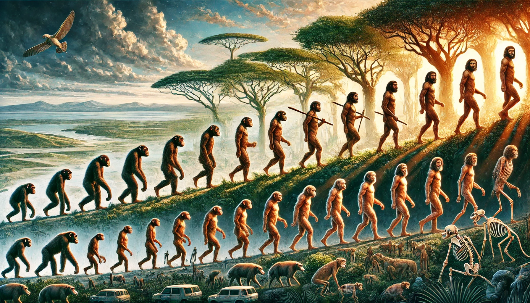
The human body presents a compelling narrative of evolution, encapsulated through millions of years of adaptation and phylogenetic change. From skeletal elements that facilitated arboreal lifestyles to organs that have lost their primary function, our anatomy is replete with vestiges of evolutionary transitions that have culminated in the present form of Homo sapiens. In addition to these residual features, evidence of ongoing evolutionary dynamics within our species is readily apparent. This article delves into these vestigial structures, such as bones and organs, and explores contemporary examples of human evolution, illuminating the continuous impact of evolutionary forces on the human phenotype.
Vestigial Bones: Echoes of a Distant Phylogenetic Heritage
One of the most salient examples of vestigial structures in the human body is the coccyx, or tailbone. The coccyx consists of a set of fused vertebrae at the base of the spine, representing the vestige of an ancestral tail. This structure harkens back to a time when early hominins and their primate predecessors possessed tails that assisted in maintaining balance and arboreal navigation. Though modern humans no longer require a tail for postural balance, the coccyx remains as an anatomical relic that underscores our shared lineage with other primates (Straus, 1944).
Another significant vestigial feature is the appendix. Historically deemed a useless remnant, the appendix is thought to have played a pivotal role in the digestion of cellulose in the diet of early herbivorous ancestors (Smith et al., 2013). While the appendix has largely lost this digestive role, it may retain some secondary function related to the immune system, serving as a reservoir for beneficial gut microbiota. Nonetheless, its redundancy is underscored by the fact that appendicitis—an inflammatory condition of the appendix—poses a significant health risk, suggesting that the appendix persists as an evolutionary remnant rather than a critical organ.
The third molars, commonly referred to as wisdom teeth, also exemplify vestigial structures. Early hominins had larger mandibles and a diet that necessitated extensive mastication of coarse, fibrous plant materials, which in turn required the additional molars. However, as early humans began to cook food and transition to a softer diet, the size of the human jaw reduced, rendering these third molars redundant and often problematic. Today, many individuals require surgical extraction of these molars due to insufficient space or malocclusion, further highlighting their evolutionary obsolescence.
The plica semilunaris, a small fold of tissue located at the medial canthus of the human eye, is another vestigial structure. This remnant represents the vestige of the nictitating membrane or "third eyelid"—a structure that remains functional in other vertebrates, such as birds and reptiles, to provide additional protection and moisture to the eye. In humans, the plica semilunaris serves no significant purpose, yet its presence speaks to a shared ancestry with species in which this feature retains functionality.
Vestigial Organs: Muscles and the Legacy of Evolutionary Function
The human body also retains muscles that no longer serve any discernible adaptive function. The palmaris longus muscle in the forearm, which is absent in approximately 14% of the population, provides a salient example. This muscle was likely essential for our tree-dwelling ancestors, enhancing grip strength and enabling effective climbing (Kaplan, 1984). The variability in the presence of the palmaris longus highlights the gradual diminishment of functionally obsolete structures through evolutionary processes.
The arrector pili muscles, responsible for producing goosebumps, are another vestigial feature. In our mammalian ancestors, these muscles facilitated piloerection, making the body appear larger in the face of threats or improving thermal insulation by trapping air. As humans lost the dense body hair characteristic of their ancestors, the function of the arrector pili muscles became largely redundant, though they still contract reflexively in response to cold or emotional stimuli (Darwin, 1872).
Jacobson's organ, or the vomeronasal organ (VNO), is another vestigial remnant in humans. In many animals, the VNO is integral to detecting pheromones, thus playing a critical role in social and reproductive behaviors. In humans, this organ is present during fetal development but regresses and becomes vestigial in adults. Although some debate persists regarding its residual presence in humans, there is little evidence to suggest that it contributes to chemosensory perception.
The plantaris muscle, located in the lower leg, is yet another vestigial structure. Evolutionary biologists suggest that this muscle was once involved in grasping, much like the feet of non-human primates that are used for climbing. Today, the plantaris muscle is functionally insignificant and can even be absent in certain individuals without any detriment to their overall mobility or muscular function.
Additional Evidence of Human Evolutionary Trajectories
Beyond vestigial structures, other facets of human biology provide compelling evidence of evolutionary processes. One example is the phenomenon of atavisms—ancestral traits that sporadically reappear in modern humans. Atavisms are rare but offer remarkable insights into evolutionary history. For instance, some individuals are born with an extended tail-like structure, a direct atavistic reflection of our primate ancestors that possessed fully developed tails (Hall, 1984).
The morphology of the human foot is another testament to our evolutionary journey from arboreal locomotion to bipedalism. Our distant ancestors possessed feet similar to those of extant apes, characterized by an opposable hallux (big toe) that facilitated climbing. As humans adopted an upright gait, the foot evolved to become a more stable structure capable of bearing weight, with arches that provide mechanical support and alignment conducive to bipedal locomotion (Latimer & Lovejoy, 1989).
The evolution of the human cranium is a particularly salient piece of evidence. Early hominins, such as Australopithecus, exhibited smaller cranial capacities, prominent supraorbital ridges, and larger jaws adapted for a robust diet. Over evolutionary time, the human cranium expanded to accommodate a significantly larger brain, and the facial structure became more gracile. These changes reflect the growing reliance on complex cognition and the reduced necessity for large masticatory muscles, especially with the development of tools and cooking methods that facilitated a softer diet (Wood & Collard, 1999).
Genomic evidence further substantiates the theory of human evolution. Comparisons between the human genome and those of other great apes, such as chimpanzees, reveal a genetic similarity of approximately 98-99%, pointing toward a shared common ancestor (King & Wilson, 1975). Additionally, the presence of endogenous retroviruses (ERVs) in the human genome offers compelling evidence of common ancestry. ERVs are viral elements that became integrated into the germline DNA of our ancestors and are inherited across generations. The fact that humans and other primates share a significant number of these viral sequences at homologous loci provides strong evidence of a shared evolutionary history (Bannert & Kurth, 2004).
Ongoing Evolution in the Human Species
While vestigial structures are emblematic of our evolutionary past, human evolution remains an ongoing phenomenon. Recent genetic studies have highlighted several adaptations that continue to shape the human species. One prominent example is lactase persistence. Historically, most humans experienced a decline in lactase production after weaning, leading to lactose intolerance in adulthood. However, in populations that domesticated dairy animals, a genetic mutation emerged that enabled continued lactase production into adulthood, facilitating the digestion of lactose (Bersaglieri et al., 2004). This adaptation is particularly prevalent among populations of European descent, underscoring the role of cultural practices in shaping genetic evolution.
Another example of ongoing evolution is the persistence of the sickle cell allele in certain populations. In regions of sub-Saharan Africa where malaria is endemic, the sickle cell trait provides a protective advantage against malarial infection. While homozygosity for the sickle cell allele results in sickle cell disease, heterozygous individuals experience increased resistance to malaria, demonstrating a case of balancing selection that continues to influence the genetic makeup of affected populations (Allison, 1954).
Moreover, recent studies have suggested changes in the human vascular system. The median artery, an artery in the forearm that typically regresses during fetal development, is now observed in approximately one-third of the population. This persistence may reflect ongoing evolutionary adaptations that favor increased blood supply to the hand, possibly in response to changes in manual dexterity and activity patterns (Lucas et al., 2020).
Conclusion
The evidence for evolution within the human species is inscribed in both vestiges of our ancestral past and ongoing adaptations. Structures like the coccyx and muscles such as the palmaris longus and arrector pili serve as enduring reminders of our evolutionary origins, while organs like the appendix and wisdom teeth illustrate the gradual obsolescence of once-crucial features. At the same time, contemporary adaptations, including lactase persistence and resistance to infectious diseases, underscore the dynamic nature of human evolution in response to shifting environmental and cultural pressures. Evolution is not a relic of the past but an ongoing force that continues to shape the human species—an enduring process reflected in our anatomy, our genetics, and our interactions with the world.
References
-
Allison, A. C. (1954). Protection Afforded by Sickle-cell Trait Against Subtertian Malarial Infection. British Medical Journal, 1(4857), 290-294.
-
Bannert, N., & Kurth, R. (2004). Retroelements and the human genome: New perspectives on an old relation. Proceedings of the National Academy of Sciences, 101(suppl 2), 14572-14579.
-
Bersaglieri, T., Sabeti, P. C., Patterson, N., Vanderploeg, T., Schaffner, S. F., Drake, J. A., ... & Lander, E. S. (2004). Genetic Signatures of Strong Recent Positive Selection at the Lactase Gene. The American Journal of Human Genetics, 74(6), 1111-1120.
-
Darwin, C. (1872). The Expression of the Emotions in Man and Animals. John Murray.
-
Hall, B. K. (1984). Developmental mechanisms underlying the formation of atavisms. Biological Reviews, 59(1), 89-124.
-
Kaplan, E. B. (1984). The Embryology of the Palmaris Longus Muscle. Journal of Hand Surgery, 9(2), 243-247.
-
King, M. C., & Wilson, A. C. (1975). Evolution at two levels in humans and chimpanzees. Science, 188(4184), 107-116.
-
Latimer, B., & Lovejoy, C. O. (1989). The calcaneus of Australopithecus afarensis and its implications for the evolution of bipedality. American Journal of Physical Anthropology, 78(3), 369-386.
-
Lucas, T., Stolarz, A., & Sharkey, J. (2020). Persistence of the Median Artery in Humans: A Modern Frequency Study. Journal of Anatomy, 237(4), 651-656.
-
Smith, H. F., Parker, W., Kotzé, S. H., & Laurin, M. (2013). Multiple Independent Events of Appendix Evolution and Related Ecological Correlations. Comptes Rendus Palevol, 12(4), 339-354.
-
Straus, W. L. (1944). The Human Tail: A Rare Vestigial Structure. The Quarterly Review of Biology, 19(1), 1-26.
-
Wood, B., & Collard, M. (1999). The human genus. Science, 284(5411), 65-71.
Author
Lander Compton
Creation Date
19:07 at 10/23/2024
Last Updated
22:47 at 10/31/2024